

A research team led by Li Rihui, assistant professor in the Centre for Cognitive and Brain Sciences at the University of Macau (UM), in collaboration with a team led by Xu Qiong, professor in the Children’s Hospital of Fudan University, has published two related studies in Molecular Psychiatry, a leading journal in psychiatry. Together, the two back-to-back publications systematically reveal the distinct and shared neural substrates of idiopathic autism spectrum disorder (ASD) and fragile X syndrome (FXS)—the most common single-gene genetic subtype of ASD—through analyses of brain functional connectivity and anatomical structure. The findings provide important insights into the pathophysiology of these neurodevelopmental disorders and offer guidance for the development of future targeted interventions.
ASD is a highly heterogeneous neurodevelopmental disorder affecting about 1 to 2% of children globally. To address this complexity, the research teams focused on FXS—the most common monogenic cause of ASD—as a genetically more defined model, enabling a clearer investigation of the shared and distinct neural mechanisms underlying the overlapping behavioural features of ASD and FXS.
In the first study, the team used the resting-state functional MRI (fMRI) method to conduct advanced analyses of brain functional connectivity and network topology in 150 children, including 70 children with idiopathic ASD, 37 with FXS, and 43 typically developing (TD) controls. The study found a unique functional alteration in children with FXS (Figure 1). They exhibited significantly weaker intrinsic connectivity within the default mode network (DMN), which is crucial for self-referential cognition, memory, and emotional regulation. This impairment clearly distinguished FXS from both ASD and TD children. At the same time, both ASD and FXS groups showed weaker connections within the cerebellar network (CN), and weaker connections between the CN and other key networks, including the DMN, sensorimotor, and visual network, compared to TD controls. These findings suggest cerebellar dysfunction as a shared neural substrate underlying core behavioural phenotypes. The study also identified critical correlations between brain networks and core symptoms of ASD (Figure 2). The relationship between the severity of core symptoms (social affect and restricted repetitive behaviour) and connectivity between several networks differed for FXS versus ASD.
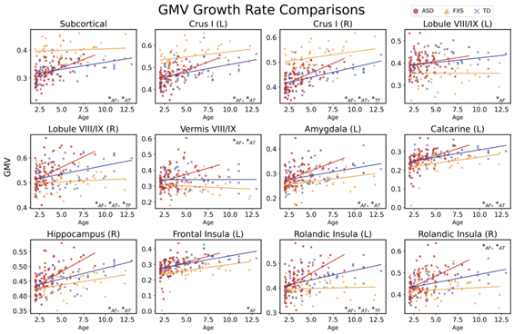
The second study focused on brain anatomy. The team used T1-weighted structural MRI data from 190 children, including 90 children with idiopathic ASD, 46 with FXS, and 54 TD controls to analyse grey matter volume (GMV) and its developmental trajectories. The findings revealed a pronounced and distinct neuroanatomical signature for FXS in early childhood (Figure 3). Compared with both idiopathic ASD and TD groups, children with FXS exhibited a complex pattern of regional volume alterations: significantly increased GMV in subcortical structures (most notably the caudate nucleus) and cerebellar Crus I, alongside substantial volume reductions in key cortical and cerebellar regions like the frontal insular regions and cerebellar vermis. This combination of overgrowth and undergrowth constitutes a clear structural ‘fingerprint’ for FXS. Importantly, these structural differences were dynamic and age-dependent (Figure 4). The GMV alterations in FXS remained consistent across narrow age ranges between two and eight years. The pivotal discovery was the significant divergence in GMV growth rates between the two disorders. Children with idiopathic ASD demonstrated a pattern of significantly accelerated GMV growth across the whole brain and nearly all regions of interest compared to both FXS and TD groups (Figure 5). In contrast, the FXS group showed growth trajectories either like TD children or significantly slower than the idiopathic ASD group in certain cerebellar regions. These results identify aberrantly accelerated brain growth as a key feature of idiopathic ASD in early childhood, distinguishing it from the developmental path of FXS.
Taken together, the two studies provide a detailed map of early brain development in ASD and FXS from both functional and structural perspectives. The first study identifies DMN hypoconnectivity as a neural signature of FXS and highlights cerebellar dysfunction as a shared pathological foundation, while the second study delineates a specific structural fingerprint for FXS and reveals aberrantly accelerated brain growth as a characteristic of ASD. Collectively, the findings support a move beyond behaviour-based diagnostic frameworks towards mechanism-driven, targeted interventions tailored to the specific neurodevelopmental trajectory of different subtypes of ASD.
Li and Xu are co-corresponding authors of both studies. In the first study, Qiao Zhongwei, professor in the Children’s Hospital of Fudan University also serves as a co-corresponding author. UM doctoral student Feng Danyong and Li Dongyun, a physician at the Children’s Hospital of Fudan University, are co-first authors of both studies. The studies were supported by the National Natural Science Foundation of China (File Nos: 82171540, 82301743), the Science and Technology Development Fund of the Macao SAR (File Nos: 0010/2023/ITP1, 0016/2024/RIB1), UM (File Nos: SRG2023-00015-ICI, MYRG-GRG2024-00296-ICI, MYRG-CRG2024-00022-ICI), the Natural Science Foundation of Anhui Province (File No: 2308085MH255), the China Medical Board (File No: 22-471), the Foreign Expert Program of the Ministry of Science and Technology (File No: G2022132004L), the Academic Leaders Development Program of the Children’s Hospital of Fudan University (File No: EKXDPY202306), and the Med+X Cross-disciplinary Team Project of the Children’s Hospital of Fudan University.
| Source: Institute of Collaborative Innovation | |
| Media Contact Information: | |
| Communications Office, University of Macau | |
| Albee Lei | Tel: (853) 8822 8004 |
| Bell Leong | Tel: (853) 8822 8009 |
| Email: | prs.media@um.edu.mo |
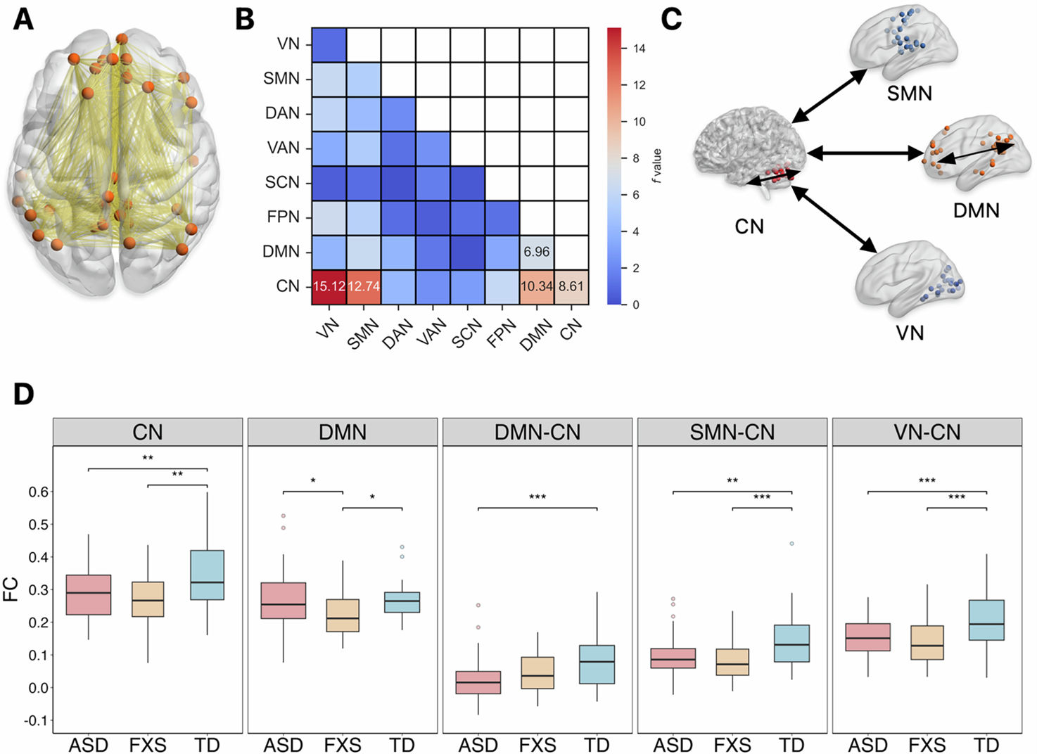
Comparisons of mean functional connectivity within and between intrinsic brain networks reveal significant group differences
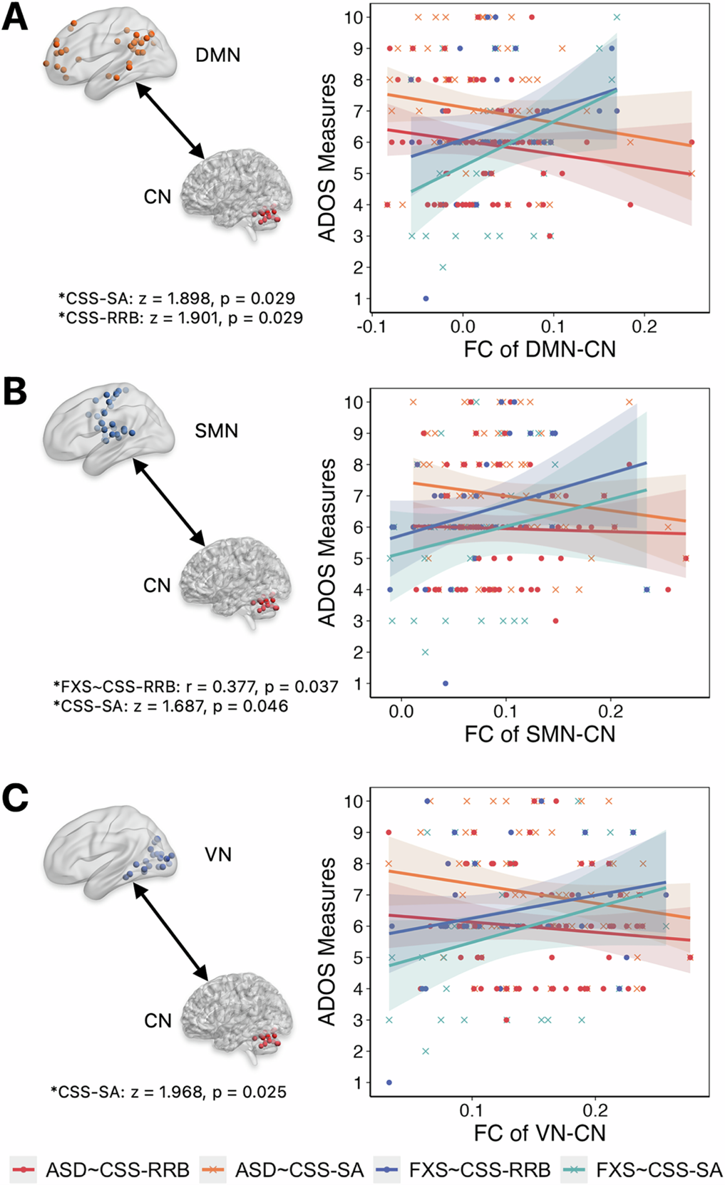
Brain network functional connectivity is correlated with multi-domain cognitive-behavioural characteristics assessed by ADOS-2
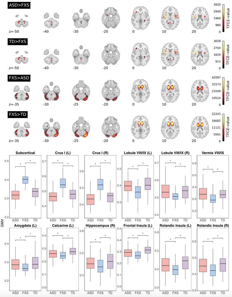
Comparisons of grey matter volume (GMV) among the three groups
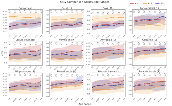
Comparisons of GMV across different age ranges among the three groups
Comparisons of GMV growth among the three groups